Evaluation of Vertebral Level of Sternal Angle and Sternal Notch Using MRI
1 Department of Anatomical Sciences, Ahvaz Jundishapur University of Medical Sciences, Ahvaz, Iran
2 Student Research Committee, Ahvaz Jundishapur University of Medical Sciences, Ahvaz, Iran
3 Department of Anatomical Sciences, Dezful University of Medical Sciences, Dezful, Iran
Citation: Bijan-Nejad D, Asadi-Fard Y, Dahaz S, Heidari-Moghadam A. Evaluation of Vertebral Level of Sternal Angle and Sternal Notch Using MRI. SF J Clin Anat Res. 2021;1(1):1001.
Abstract
Purpose: Surface anatomy and living anatomy is essential for understanding the foundation of physical examination, and the interpretation of clinical findings. The sternal angle is an important clinical landmark for identifying many other anatomical points. Many books have stated that sternal angle (Louis angle) passing of T4-T5 intervertebral disc. Numerous inconsistencies in clinically important surface markings exist between and within anatomical reference texts. The purpose of this study is to determine the vertebral level of this plane in living subjects.
Methods: MRIs of 200 patients with thoracic spine were used. The vertebral level of the sternal angle and sternal notch were determined on sagittal scans as the level at which a horizontal line through the manubriosternal joint and sternal notch intersected the anterior border of the vertebral column.
Results showed that the vertebral level of sternal angle ranged between T3-T4 intervertebral disc to T6 and the vertebral level of sternal notch ranged between T1-T2 to T3-T4 intervertebral disc.
Conclusion: It is a known fact that there is a difference in the vertebral level of various mediastinal structures between cadavers and living subjects. Thus the students and the clinicians must be aware of the changes in the vertebral level of sternal angle.
Keywords: Surface anatomy; Sternal angle; Sternal notch; MRI
Introduction
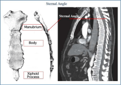
Surface anatomy is an essential component of studying the human body [1]. It is not just about knowing what lies under the skin and what structures are perceptible to touch in a living body, it is also about enabling learners to improve their skills in clinical examinations, interventional procedures [1] and interpretation of diagnosing images. Surface anatomy and living anatomy is also essential for understanding the foundation of physical examination, and the interpretation of clinical findings. The sternum is easily palpable and is comprised of the manubrium, body and xiphoid process. The manubrium lies superiorly with the suprasternal or jugular notch marking its upper border, which is easily palpable between the clavicular heads. The upper border of the manubrium is used as a landmark when making a mediastinoscopy or collar incision. It also corresponds to the level of the lower border of the second thoracic vertebra and first thoracic spinous process. The manubrium is 4 cm long and overlies the aortic arch [2]. The manubrium articulates with the sternal body at the angle of Louis: this manubriosternal junction is palpable as a transverse ridge in most patients. The second costal cartilage articulates with the lateral border of the sternum at the angle of Louis (Figure 1). Counting the ribs anteriorly is most easily started at this level because the first rib is impalpable beneath the clavicle. The angle of Louis lies at the level of the lower border of the fourth thoracic vertebra [2]. The sternal body is 10 cm long and lies opposite the fifth to eighth vertebrae in front of the heart. Below the body is the cartilaginous xiphoid, which may be palpable, and lies at the level of the ninth thoracic vertebra. The plan passing through the angle of Louis and the lower border of the fourth thoracic vertebra is an important landmark for deeper thoracic structures. Mediastinal structures said to lie on the Louis plane include the bifurcation of trachea, the concavity of arch of aorta and the azygos arch. To obtain a rational and convenient method of subdividing the trunk, anatomists have traditionally used a number of vertical and horizontal lines. Also Surgical approach to the upper thoracic spine has always been challenging [3,4]. It involves major procedures, such as sternotomy and thoracotomy, which are related with high morbidity
and mortality. It was recently shown that, on midsagittal thoracic Magnetic Resonance Imaging (MRI) scan, a tangential line drawn on the upper limit of the sternal notch and then extended bisected the upper thoracic spine to a level up to the lower border of T4 in some cases and up to T3 in most cases [4]. It was also illustrated that such a line represents the level of upper thoracic vertebra accessible with a low anterior cervical approach without sternotomy or thoracotomy. Anatomical planes used in clinical practice and teaching anatomy are largely derived from cadaver studies. Numerous inconsistencies in clinically important surface markings exist between and within anatomical reference texts. The aim of this study was to reassess the accuracy of common anatomical planes in vivo using magnetic resonance imaging. The purpose of this study is to determine the vertebral level of this plane in living subjects.
Materials and Methods
This descriptive study was performed in the Kianpars Imaging center, Ahvaz, Iran. MRIs of 200 patients with thoracic spine were used. In this study, individuals with different groups were present in the range of 20 to 60 years. The thoracic MRI was done with a Signa unit (General Electric, USA) operating at field strength of 3 Tesla. T1 weighted sagittal, T2 weighted sagittal were obtained using a spin echo pulse Sequence. The slice thickness was 4 mm for sagittal images. Images of selected subjects were recalled on the monitor and sections include complete thoracic spine and sternum selected and highlighting the sternal angle and sternal notch. At MR imaging, the sternal anatomy is best depicted with T1-weighted spin-echo pulse sequences. Age and gender were available in all cases. For the numbering of vertebrae first we performed T2 weighted sagittal
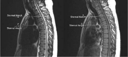
from cervical vertebra and numebring performed from C3. The vertebral level of the sternal angle and sternal notch were determined on sagittal scans as the level at which a horizontal line through the manubriosternal joint and sternal notch intersected the anterior border of the vertebral column. The plane was recorded according to whether it intersected the upper half of a vertebral body, lower half, or the intervertebral disc (Figure 2). The patient breathed slowly until the breathing movements did not cause excessive sternum movement.
Result
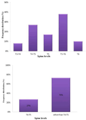
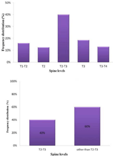
The vertebral level of sternal angle ranged between T3-T4 intervertebral disc to T6. The sternal angle passed through T4-T5 intervertebral disc in 54 case (27%) and through other level in 146 case (73%). T5-T6 intervertebral disc is the commonest vertebral level of sternal angle in present study (76 case, 38%) (Figure 3). The vertebral level of sternal notch ranged between T1-T2 to T3-T4 intervertebral disc. The sternal angle passed through T2-T3 intervertebral disc in 80 cases (40%) and other level in 120 cases (60%) (Figure 4). T2-T3 level is the commonest vertebral level of sternal notch in our study. Analysis of variance showed no significant correlation (age vs. level: P=0.087, sex vs. level: P=0.061).
Discussion
There are many surface anatomical symptoms that have clinical implications. These surface symptoms are guide for achieving the inner organs, vessels and nerve. These surface symptoms can indicate the place of start, end, or division of a vessel. For example, immediately below the inguinal ligament is a good way to achieve
femoral artery for catheterization. The surface sign that weexamined in this study using MRI includes the sternal angle and the sternal notch. The angle at the junction of the manubrium and the body of the sternum (sternal angle) bears the eponymous name “angle of Louis”. Pierre Charles Alexandre Louis (1787-1872), a French lawyer turned physician. If we draw horizontal line from this angle to the spine, by this line the mediastinum is divided into two upper and lower parts which gives us information about the elements of the mediastinum. If we draw tangential line from on the upper limit of the sternal to spine probably bisected the upper thoracic spine to a level up to the lower border of T4 in some cases and up to T3 in most cases which is used for surgical approach to the upper thoracic spine. The variations of the sternal angle level and sternal notch level observed in our study.
Chukwuemeka et al., from London in their study of CT chest on 51 subjects found that the vertebral level of sternal angle was T4 in 18.6% cases, T5 in 72.5% cases and T6 in 7.9% cases [5]. Syed Naziya P et al., in their study of CT chest on 48 subjects showed that study the vertebral level of sternal angle pass through T4 and above in 22.91% cases, T5 in 68.75% cases and T6 in 8.33% cases [6]. In the present study the vertebral level of sternal angle was found to pass through T4-T5 level in 54 cases (27%) and T5-T6 level in 76 cases (38%). These values when compared indicated that the average value of the vertebral level of sternal angle found in present study was very similar to that found by Chukwuemeka et al [5]. The commonest vertebral level found in present the study is T5-T6 level, which is one vertebra lower than the classically described level of sternal angle in cadaveric dissections. It is a known fact that there is a difference in the vertebral level of various mediastinal structures between cadavers and living subjects. Thus the students and the clinicians must be aware of the changes in the vertebral level of sternal angle.
Acknowledgment
We thank Student Research Committee of Ahvaz Jundishapur University of Medical Sciences for supporting this study under agreement number 97S46, in 2019.
Author Contribution
DBN: Manuscript writing. AHM: Data management, data analysis. SHD: Clinical consult. YAF: Data analysis, manuscript editing.
References
- Leonard RJ, Acland RD, Cahill DR, Casey MA, Ger R, Gilroy AM, et al. The clinical anatomy of several invasive procedures. Clin Anat. 1999; 12: 43-54.
- Standring S. Gray’s Anatomy. The Anatomical Basis of Clinical Practice. Churchill Livingstone. 2000.
- Capener N. The evolution of rhachotomy. J Bone Joint Surg Br. 1954; 36: 173-179.
- Sharan AD, Przybylski GJ, Tartaglino L. Approaching the upper thoracic vertebrae without sternotomy or thoracotomy: a radiographic analysis with clinical application. Spine. 2000; 25: 910-916.
- Chukwuemeka A, Currie L, Ellis H. CT anatomy of the mediastinalstructures at the level of the manubriosternal angle. Clin Anat. 1997; 10: 405-408.
- Syed N, Althaf A, Pritha S, Syed I, Anand A, Murthy G. A Computerized Tomographic Study of Normal Mediastinal Anatomy in Indian Subjects. International Journal of Health Sciences &Research. 2014; 4: 111-116.
Figures