Placental Chorioangioma – A Benign Vascular Tumor of Placenta
Department of Radio Diagnosis, Dr. VVPF Medical College, Maharashtra University of Health Sciences, India
Citation: Patel DK, Kachewar SG. Placental Chorioangioma – A Benign Vascular Tumor of Placenta. SF J Radiol Clin Diagn. 2021;3(1):1007.
Abstract
Chorioangiomas are the most common placental tumors. They are benign vascular tumors. They are usually detected incidentally on ultrasonography with most of them being asymptomatic. Tumors of more than 4 cm usually are associated with fetal and maternal complications. In this article we present a case of Chorioangioma which was detected in second trimester of pregnancy associated with only Single Umbilical artery. It was associated with polyhydramnios causing premature rupture of membranes leading to a premature delivery at 25 weeks. Newborn did not survive post-delivery. Placenta was removed with tumor and diagnosis was confirmed.
Keywords: Chorioangioma; Single umbilical artery; Polyhydramnios
Case Presentation
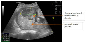
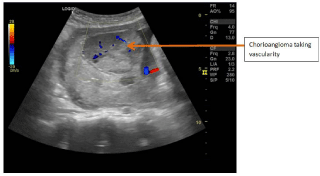
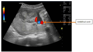
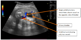
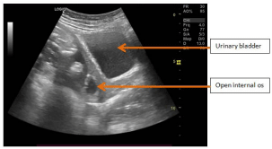
A 21 year old female presented at 27th week of gestation with complaints of per vaginal leaking since last 24 hours. On examination frank per vaginal leaking was noted. Patient was then sent for Ultrasonography. On Ultrasonography, Fetal gestational parameters were of 25 weeks 2 days. Estimated weight of baby was 873 gms. Placenta was posterior in location. A well-defined 53x43x58 mm (MLxAPxCC) heterogeneously hypoechoic to isoechoic lesion was noted on the fetal surface of the placenta, at the fundal end (Figure 1). Doppler examination showed vascularity, with low RI (Figure 2). Umbilical cord was seen inserted adjacent to the inferior border of lesion (Figure 3) and it showed only two vessels, one umbilical vein and one umbilical artery (Figure 4). Fetus was noted in breech position with fetal head and neck near the lesion. Internal Os was open (Figure 5). AFI was 11 cm. Above
Radiolgical features were s/o chorioangioma of Placenta.
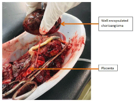
Delivery was inevitable and Pregnancy was terminated by normal vaginal delivery. Unfortunetly infant did not survived. Placenta showed a well-defined encapsulated lesion which was easily separated from placenta (Figure 6).
Discussion
Chorioangioma is a benign placental tumour, defined by the abnormal proliferation of vessels arising from chorionic tissue [1]. Giant choriangiomas are rare placental tumours, associated with a high prevalence of pregnancy complications and a poor perinatal outcome [2]. Prenatal diagnosis of chorioangioma is achieved by ultrasonography with colour Doppler [3].
These tumors act as large arteriovenous shunts within the placenta, diverting blood away from the fetus. Polyhydramnios has been linked to increased urine production, associated with fetal hyperdynamic circulation related to shunting of blood or fetal anaemia. It has also been proposed that transudation of fluid from the tumour surface contributes to accumulation of amniotic fluid [4].
Chorioangiomas are usually found on the fetal surface of the placenta, often in the vicinity of umbilical cord insertion. Grossly they are well-circumscribed, purplish red tumours with fleshy, congested, red to tan cut surface [5].
The presented case was unique which showed Chorioangioma which was more than 4 cm in diameter and was asscociated with single umbilical artery. Mother developed polyhydramnios which is a known complication, which further led to premature labour and inevitable delivery.
References
- Wallenburg HC. Chorioangioma of the placenta. Thirteen new cases and a review of the literature from 1939 to 1970 with special reference to the clinical complications. Obstet Gynecol Surv. 1971; 26: 411-425
- Zanardini C, Papageorghiou A, Bhide A, Thilaganathan B. Giant placental chorioangioma: natural history and pregnancy outcome. Ultrasound Obstet Gynecol. 2010; 35: 332-336.
- Prapas N, Liang RI, Hunter D, Copel JA, Lu LC, Pazkash V, et al. Color Doppler imaging of placental masses: differential diagnosis and fetal outcome. Ultrasound Obstet Gynecol. 2000; 16: 559-563.
- Isaacs H Jr. Fetal hydrops associated with tumors. Am J Perinatol. 2008; 25: 43-68.
- Barros A, Freitas AC, Cabral AJ, Camacho MC, Costa E, Leitao H, et al. Giant placental chorioangioma: a rare cause of fetal hydrops. BMJ Case Reports. 2011; 02: 3880.
Figures